
Knee Conditions
& Treatment
Knee Overview
Whether you're pushing through miles on the road, cutting on the field, or just trying to keep up with your kids, your knees absorb more impact than almost any other joint in your body. Dr. Eskew is a fellowship-trained knee surgeon who understands that knee pain can affect athletes at any level and any age, and that it's often more disruptive than people expect. The good news is that most knee pain responds well to conservative treatment, such as anti-inflammatories, targeted therapeutic exercise, and activity modification.
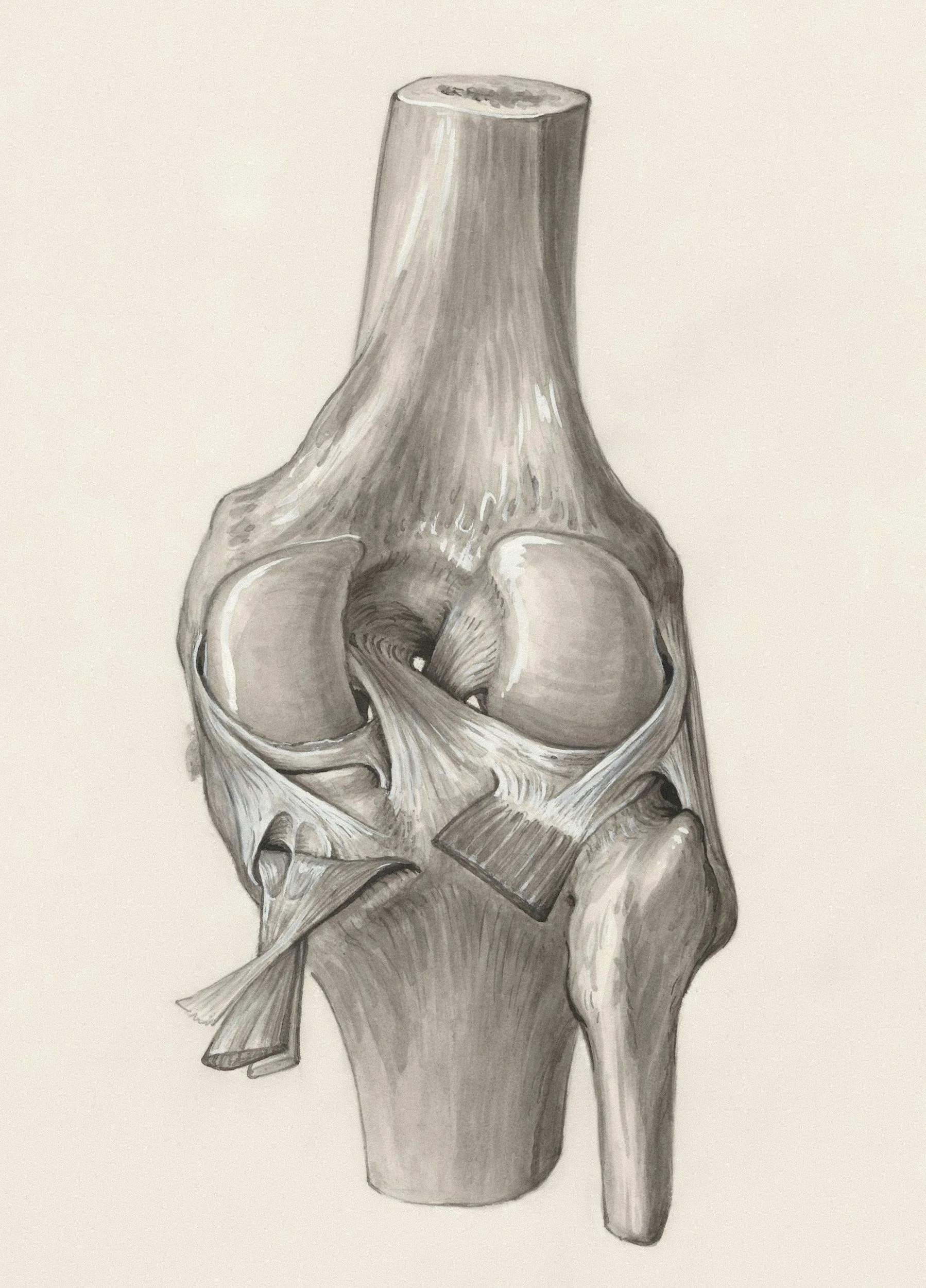
The knee is more than a simple hinge. It's made up of three compartments where the femur, tibia, and kneecap meet, cushioned by cartilage and the meniscus, which absorb impact and allow smooth movement with every step, stride, or landing. Holding it all together is a network of ligaments, including the ACL, PCL, MCL, and LCL, that stabilize the joint through cutting, pivoting, and rapid changes of direction. When any one of these structures is injured or wears down over time, it can throw off the entire joint, which is why Dr. Eskew starts with an accurate diagnosis of exactly which part of the knee is involved.
Common Knee Conditions
Because the knee gets little stability from bone alone, it depends heavily on its ligaments and surrounding muscles to stay stable through movement. This is part of why certain injuries call for a closer look, and Dr. Eskew may recommend an MRI to see exactly what's happening beneath the surface. When a ligament or cartilage tear is found, arthroscopic knee surgery may be the right next step to repair it and get you back to moving with confidence.
-
The meniscus is a shock-absorbing cushion between the thigh bone and shin bone, and it's one of the most commonly injured structures in the knee. Tears can happen suddenly from twisting or pivoting, or gradually from wear over time.
Common signs and symptoms include:
Pain along the joint line
Swelling that builds over hours
Catching, locking, or the knee giving way
Difficulty fully straightening or bending the knee
Treatment options:
Nonoperative care. Many tears, especially degenerative ones, improve with physical therapy and activity modification.
Meniscectomy. When a tear isn't repairable, removing the damaged portion offers fast, reliable relief.
Meniscal repair. Whenever possible, repair is favored to preserve the meniscus and protect the knee from early arthritis. Techniques include inside-out, outside-in, and all-inside repair, chosen based on the tear's location.
Root tears, bucket handle tears, and LMORTs
Some tear patterns are treated differently. Root tears occur where the meniscus attaches to bone. Once that attachment fails, the meniscus can no longer function as a shock absorber, so root tears are treated as a more urgent surgical priority to protect the knee. Bucket handle tears and lateral meniscus oblique radial tears (LMORTs) are large, often severe tear patterns strongly linked to trauma and frequently found alongside ACL injuries, since the same twisting injury often damages both structures together.
Dr. Eskew evaluates your full knee, including any associated ligament injury, to determine the right treatment and timing for your specific tear.
-
The anterior cruciate ligament (ACL) is one of the primary stabilizing ligaments of the knee, controlling rotation and forward movement of the shinbone. ACL tears usually happen suddenly, often during cutting, pivoting, or landing from a jump, and are among the most common serious knee injuries in sports.
Common signs and symptoms include:
A pop felt or heard at the moment of injury
Immediate swelling
A sense of instability or the knee giving way
Difficulty continuing activity right after injury
Pain and limited range of motion in the days that follow
Not every ACL tear requires surgery. Lower-demand patients with a stable knee may do well with physical therapy and activity modification alone, though nonoperative treatment carries a higher risk of ongoing instability, particularly for athletes returning to cutting and pivoting sports.
When surgery is the right choice, Dr. Eskew offers the following graft and technique options, tailored to your anatomy, activity level, and goals:
Bone-patellar tendon-bone (BPTB) autograft: known for fast, reliable bony healing and among the lowest revision rates of any graft option
Hamstring tendon autograft: a smaller incision with a quick recovery, a strong option for many active patients
Quadriceps tendon autograft: an increasingly popular choice known for excellent strength and reliable outcomes
Lateral extra-articular tenodesis (LET): an additional procedure frequently performed alongside ACL reconstruction in Dr. Eskew's practice to add rotational stability and meaningfully reduce the risk of re-tear, especially valuable for higher-risk patients
Graft choice is only part of the picture. Every knee is different, and Dr. Eskew evaluates factors that many surgeons overlook, including your posterior tibial slope (the natural angle of your shinbone, which can influence graft stress and re-tear risk), whether you have generalized ligamentous laxity, and sex-specific differences in injury patterns, since female athletes face a meaningfully higher rate of ACL injury and warrant tailored consideration in both graft choice and technique.
Because no two ACL injuries or knees are exactly alike, Dr. Eskew builds a reconstruction plan around your specific anatomy and risk factors, not a one-size-fits-all protocol, to give you the most durable path back to the sport you love.
-
Cartilage covers the ends of the bones in the knee, allowing smooth, pain-free movement. When cartilage is damaged, whether from an acute injury or osteochondritis dissecans (OCD), a condition where a piece of bone and cartilage loses its blood supply and can separate from the joint, the result is often pain, swelling, and mechanical symptoms that don't resolve on their own.
Common signs and symptoms include:
Pain, especially with activity or weight-bearing
Swelling that recurs with certain movements
Catching, locking, or a sense of instability in the knee
In OCD, symptoms that may be vague or intermittent before a fragment becomes unstable
Treatment starts with understanding the full picture, not just the cartilage defect itself. Knee alignment plays a major role in how much stress is placed on the damaged area, and correcting significant malalignment is often an essential part of giving any cartilage treatment the best chance to succeed. Depending on the size, location, and depth of your injury, Dr. Eskew offers both whole tissue and cell-based treatment options:
Whole Tissue Options
OATS (osteochondral autograft transplantation), transferring healthy bone and cartilage from another part of your own knee
OCA (osteochondral allograft), using donor bone and cartilage to restore larger defects
Cell-Based Options
MACI (matrix-induced autologous chondrocyte implantation), growing your own cartilage cells in a lab and implanting them to regenerate the damaged surface
Particulated juvenile cartilage, using donor cartilage tissue to help fill and restore smaller defects
Autologous minced cartilage, using small fragments of your own healthy cartilage, harvested and implanted in a single procedure to help fill smaller defects
Because cartilage injuries rarely exist in isolation, Dr. Eskew evaluates your entire knee, including alignment, ligament stability, and meniscus health, to build a treatment plan that addresses the underlying cause, not just the injury itself, giving your knee the best chance at a lasting recovery.
-
A kneecap that slips out of place, even once, can leave you afraid it will happen again. That fear alone can change how you move, how you play, and how much you trust your own knee. Getting a full, honest evaluation after a dislocation matters, because treating it well the first time gives you the best shot at never dealing with it again.
Common signs and symptoms include:
A visible or felt shift of the kneecap out of place
Sudden pain and swelling after a twisting injury
A sense of the kneecap slipping, buckling, or giving way
Ongoing apprehension with pivoting or cutting movements
Here's what a lot of patients don't realize: not every first-time dislocation should be treated the same way. Some knees have underlying anatomy, a shallow trochlear groove (trochlear dysplasia), a tilted kneecap, or a shin bone attachment point that's pulling the kneecap off track (measured as the TT-TG distance) that make repeat dislocation far more likely. A dislocation can also chip off a piece of bone and cartilage on its way out (an osteochondral fracture), which changes the treatment picture entirely. In these situations, surgery after the very first dislocation is often the smarter path, rather than waiting through months of therapy that's unlikely to hold.
When surgery is the right call, MPFL reconstruction rebuilds the ligament that keeps your kneecap tracking properly, and outcomes are strong: most patients return to sport with a very low chance of dislocating again. In knees with more significant anatomic risk factors, Dr. Eskew may recommend addressing those factors directly alongside the ligament reconstruction, since skipping that step is one of the most common reasons instability comes back.
Dr. Eskew looks at your whole knee, its alignment, its anatomy, and how it moves, not just the dislocation itself, to figure out exactly why your kneecap came out and build a plan that keeps it in place for good.
-
That nagging ache just below or above your kneecap that flares up every time you jump, squat, or run stairs can feel like it's never going to fully settle down, especially when rest alone doesn't seem to touch it. The good news is that this is one of the most treatable conditions in sports medicine, and the right approach rarely involves surgery.
Common signs and symptoms include:
Pain just below the kneecap (patellar tendinitis, often called "jumper's knee") or just above it (quadriceps tendinitis)
Pain that worsens with jumping, squatting, or going up and down stairs
Stiffness after periods of rest, especially first thing in the morning
Tenderness directly over the tendon
Here's what a lot of patients don't realize: the answer to tendinitis usually isn't rest, it's the right kind of loading. Tendons respond and get stronger when they're challenged in a specific, progressive way, and a structured physical therapy program built around eccentric and single-leg exercises is the most well-supported treatment available. Eccentric loading, controlled lengthening of the tendon under tension, has consistently shown some of the best outcomes for both patellar and quadriceps tendinitis, and single-leg plyometrics help rebuild the tendon's ability to absorb the explosive, repetitive loads of jumping and cutting sports. This isn't a quick fix. It takes weeks of consistent, progressive work, but it addresses the actual problem rather than just quieting the pain temporarily.
For the small number of patients who don't improve with a genuine course of targeted therapy, additional options exist, but surgery is rarely the first or even the second step.
Dr. Eskew builds your rehabilitation program around your specific sport and demands, so you're not just pain-free at rest, but ready to jump, cut, and land with confidence again.
-
A serious knee injury can happen in a split second, a fall, a collision, a car accident, but living with the uncertainty afterward can feel like it stretches on forever. Will the knee ever feel stable again? Will you walk normally, let alone get back to sport?
Common signs and symptoms include:
Significant pain, swelling, and bruising after a fall or impact
Visible deformity or an inability to bear weight
A knee that feels loose, unstable, or shifts out of place
An inability to straighten the leg or hold it up against gravity, which can signal a tendon rupture
Numbness, a cold foot, or a weak pulse below the knee, which can signal a more serious injury needing urgent attention
Knee trauma covers a range of injuries, and getting the right diagnosis quickly matters:
Multiligament knee injuries (knee dislocation): often involving the ACL, PCL, MCL, and LCL together, this is a far more serious injury than a kneecap dislocation, even if the knee looks like it "popped back into place" on its own
Patella fractures: breaks in the kneecap, which can occur alone or alongside ligament or tendon injury
Patellar and quadriceps tendon ruptures: sudden tears of the tendons above or below the kneecap, often from a forceful landing or a fall, causing an inability to straighten or support the leg
Vascular and nerve injury: knee dislocations in particular can put nearby blood vessels and nerves at risk, which is why urgent, careful evaluation in the first hours matters
Treatment depends entirely on what's injured and how severely. Tendon ruptures generally require prompt surgical repair to restore the ability to walk and straighten the leg normally, and outcomes are best when repair happens early, before the tendon has a chance to retract and scar. Patella fractures may be treated without surgery if stable, or with open reduction and internal fixation (ORIF) if displaced. When multiple ligaments are torn, the timing and sequence of treatment matters, and a coordinated approach gives the knee its best chance at full stability.
Dr. Eskew evaluates the complete injury, bone, tendons, ligaments, nerves, and blood vessels together, not just the most obvious piece, to build a treatment plan and sequence that gives your knee the best possible path back to full function.