
Complex Cases
& Revision Surgery

Complex Cases and Revision Surgery Overview
Complex and Revision Surgery
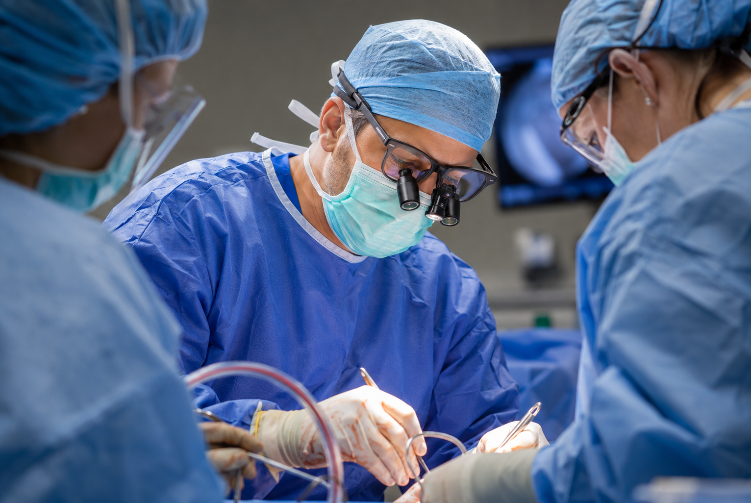
Not every case is straightforward, and not every patient is starting fresh. If a prior surgery didn't deliver the outcome you expected, or if you're facing a condition that's more complicated than a typical diagnosis, you need a surgeon who has the experience to figure out what went wrong and the skill to fix it. As an academic, tertiary referral practice, complex and revision cases like yours are something Dr. Eskew sees regularly, and he's committed to getting you a clear answer and real relief.
His expertise includes revision sports surgery, such as revision Tommy John (UCL) reconstruction and revision rotator cuff repair, as well as failed distal biceps repair and other procedures that didn't hold up the first time. He also treats complex shoulder and elbow conditions including paralytic shoulder, scapular winging, thoracic outlet syndrome, and nerve-related issues such as suprascapular nerve compression. For patients who need joint replacement or a revision of a prior replacement, Dr. Eskew offers shoulder and elbow arthroplasty, along with reconstructive options like tendon transfers when a standard repair isn't enough. Whatever brought you to this point, the goal is the same: a clear explanation of what's really going on, and a plan built to get it right.
Complex and Revision Surgical Conditions
As an academic, tertiary referral practice, complex and revision cases like yours are something Dr. Eskew sees regularly, and he's committed to getting you a clear answer and real relief.
-
A recurrent UCL injury after previous surgery is a different kind of setback. It's not the fear of a first injury anymore, it's watching the recovery clock start over, wondering if the answer this time will actually hold.
Common signs and symptoms include:
Recurring medial elbow pain or instability after a prior UCL reconstruction
Decreased throwing velocity or control that doesn't improve with rehab
A sense that the elbow never felt quite right after the original surgery
Why This Happens
Revision isn't always about a failed surgery. Graft attenuation over time, a new injury to the reconstructed ligament, mechanics that never fully corrected, or simply the cumulative stress of years of high-level throwing can all lead here. Understanding the real cause matters, since it shapes the right fix.
Revision UCL surgery is becoming more common. As more athletes have Tommy John surgery earlier, often as teenagers, more are living long enough in the sport to eventually need a second procedure. Rates of revision reconstruction have risen sharply over the past two decades.
The approach has evolved too. Revision once meant another full reconstruction with a new graft. Now, in the right candidates, repair with augmentation is increasingly used instead, offering strong return-to-sport rates with a notably faster recovery than a second reconstruction.
Dr. Eskew evaluates your prior surgery, your ligament's current condition, and your goals to determine the path most likely to hold this time.
-
Getting a joint replaced is a big decision, but needing a second surgery on that joint is a different experience entirely. You already know how hard the recovery was the first time, and facing it again, without yet knowing what will actually fix things, is a real weight to carry.
Common signs that a replacement needs revision include:
Pain that's returned or never fully resolved
Loss of motion, strength, or stability
A sense of looseness, clicking, or the joint shifting
Swelling, warmth, or other signs of possible infection
Why Revision Is Sometimes Needed
There are several well-established reasons a joint replacement may need revision, and none of them point to any one thing going wrong. The most common include:
Component loosening, where the implant gradually separates from the bone over time
Instability, when the joint dislocates or doesn't stay properly seated
Infection, which can develop soon after surgery or years later
Rotator cuff issues, specific to anatomic shoulder replacement, where the surrounding tendons no longer support the joint as intended
Wear or mechanical issues with the implant itself after years of use
Primary Replacement
For patients facing joint replacement for the first time, Dr. Eskew offers anatomic and reverse shoulder replacement, as well as total elbow replacement, matched to your specific condition, bone quality, and goals.
Revision Replacement
Revision surgery is more complex than a first-time replacement, working around scar tissue, altered anatomy, and sometimes compromised bone. Outcomes vary depending on the underlying cause, which is why identifying it accurately before surgery matters so much.
Dr. Eskew evaluates your prior surgery, current imaging, and overall shoulder or elbow health to build a revision plan aimed at giving your joint lasting relief.
-
A rotator cuff repair that doesn't hold can be discouraging in a way that's hard to explain to people who haven't been through it. You did the physical therapy, you protected it, and the pain came back anyway. The question isn't just "can this be fixed," it's "is there anything left to fix it with."
Common signs and symptoms include:
Pain and weakness that return after an initial period of improvement
Loss of overhead motion or strength
A sense the shoulder never fully recovered from the first surgery
Why This Happens
Re-tears can happen for a range of reasons: tissue quality, tear size, how much time passed before the first repair, or simply the biology of healing not cooperating. Understanding the pattern and quality of the remaining tendon and muscle is essential before deciding what revision surgery should actually accomplish.
Evaluating the Shoulder
Not every revision candidate is the same. Dr. Eskew evaluates factors like the ROHI score (a measure incorporating rotator cuff tear characteristics and fatty infiltration that helps predict whether a repair is likely to succeed) to determine whether the tendon is repairable at all, and if so, what technique gives it the best chance.
Treatment Options
Depending on tissue quality and tear pattern, options include:
Repair with marginal convergence, bringing torn tendon edges together to reduce tension before final fixation
Biologic tuberoplasty, reshaping the bone surface to improve tendon healing potential
Patch augmentation, reinforcing a repair with a biologic or synthetic scaffold when tissue quality is compromised
Superior capsular reconstruction (SCR), restoring stability in shoulders with an irreparable tear
Lower trapezius or latissimus dorsi tendon transfer, rerouting a healthy tendon to restore function when the native rotator cuff can't be repaired
Extensive debridement and capsular releases, addressing scar tissue and stiffness that often accompany a prior failed repair
Because every revision shoulder tells a different story, Dr. Eskew builds a plan around exactly what's left to work with, giving you the most realistic and durable path back to function.
-
Some shoulders keep coming out of place no matter how careful you are. Maybe it happened reaching overhead, maybe just rolling over in bed. When a shoulder dislocates with less and less force each time, it's usually a sign that the joint itself has run out of bone to hold it in place, not that you're doing something wrong.
Common signs and symptoms include:
Repeated dislocations, sometimes with minimal trauma
A persistent feeling of looseness or apprehension
Prior labral repair that didn't hold
Dislocations occurring with everyday movements, not just sports
Why This Happens
Each dislocation can chip away small amounts of bone from the socket or the ball of the joint. Once that bone loss becomes significant, a soft-tissue repair alone often isn't enough to hold the shoulder in place, and a bone-based procedure is needed instead.
Treatment Options
Latarjet procedure: for significant glenoid bone loss, transfers a piece of bone and its attached tendon to rebuild the socket and add a mechanical block against dislocation, with strong long-term outcomes and low recurrence rates
Arthroscopic distal tibial allograft (DTA): rebuilds the glenoid's natural shape and joint surface using donor bone, without disturbing the surrounding tendons and muscle, often used for prior failed surgery or posterior instability
Other autograft and allograft options: additional bone graft techniques chosen based on the location and severity of bone loss and your prior surgical history
Open Bankart repair with capsular shift: for patients with minimal bone loss, tightens and reattaches the torn labrum and stretched capsule to restore stability without the need for a bone graft
Dr. Eskew evaluates the amount, location, and pattern of bone loss driving your instability to determine which of these approaches gives your shoulder the best chance at lasting stability.
-
Some shoulders come loose in every direction, forward, backward, and downwards, sometimes without any real trauma at all. It's disorienting, especially when it's dismissed as "just being flexible."
Common signs and symptoms include:
Subluxation or dislocation in multiple directions, often with minimal trauma
A history of being unusually flexible or "double-jointed"
Instability that hasn't responded to typical strengthening
Why This Happens
Some patients simply have naturally loose ligaments and a stretched capsule. Others have an underlying connective tissue condition like Ehlers-Danlos syndrome (EDS), where tissue throughout the body is inherently more fragile. Recognizing EDS matters, since it changes both the treatment approach and what to expect from it.
Physical Therapy Comes First
For most patients, physical therapy is the primary treatment, not just a first step. More than half improve significantly through a focused program that retrains the shoulder's surrounding muscles to compensate for what the ligaments can't. This takes real commitment, often several months, and it matters even more here, since surgery on a hypermobile or EDS shoulder carries added risk.
When Surgery Is Needed
For patients who don't improve with therapy, options include arthroscopic capsular plication, open capsular shift, and, in EDS patients with fragile tissue, allograft-augmented repair for added durability.
Dr. Eskew evaluates your instability pattern, hypermobility, and whether EDS is contributing, to build a plan grounded in the right rehabilitation first, with surgery reserved for when it's truly needed.
-
Vague symptoms in the neck, shoulder, and arm are frustrating on their own, but even more so when they don't fit neatly into any one diagnosis. Thoracic outlet syndrome (TOS) is notoriously difficult to diagnose, and it's often missed or misattributed to something else entirely, which is why it needs to be evaluated by someone who truly understands the condition.
Common signs and symptoms include:
Pain, tension, or tightness in the neck, shoulder, and shoulder blade area
Numbness or tingling in the arm or hand, often not confined to one clear nerve pattern
Symptoms that worsen with overhead activity or repetitive arm use
Why This Happens
TOS occurs when nerves passing between the neck and arm become compressed, most often by the scalene muscles, a narrowed space near the first rib, or a tight pectoralis minor muscle.
Dr. Eskew has dedicated significant research time to TOS, including authoring review articles on the condition and collaborating with Major League Baseball's physician research community to better understand how it affects throwing athletes. That depth of study translates directly into how carefully he evaluates every patient who comes in with these symptoms.
Physical Therapy First
For nearly every patient, scapular-focused physical therapy is the starting point, retraining posture and scapular mechanics to open up the compressed space. Many patients improve significantly without surgery.
When Surgery Is Needed
For patients who don't respond to a genuine course of therapy, surgical decompression may involve pectoralis minor release, scalenectomy, and, in select cases, first rib resection, tailored to exactly where the compression is occurring.
Because TOS is so frequently misdiagnosed elsewhere, Dr. Eskew takes the time to confirm what's really going on before building your treatment plan, grounded in the depth of research he's put into understanding this condition.
-
Needing a second ACL surgery brings a different kind of weight than the first. Beyond the physical recovery, there's the question of why it happened again, and whether the next fix will actually hold.
Common signs and symptoms include:
A new injury to a previously reconstructed ACL, or gradually worsening instability without a clear injury
The knee giving way or feeling unreliable during activity
Persistent swelling or a sense the knee never fully returned to normal after the first surgery
Difficulty trusting the knee during cutting, pivoting, or high-demand activity
Why This Happens
There are several well-established reasons an ACL graft may fail, and understanding which one applies matters before deciding on a revision plan. Dr. Eskew evaluates factors including posterior tibial slope (a steep slope can place added stress on the graft), any untreated meniscus or cartilage injury, ligamentous laxity, graft type and healing, and the timeline of return to sport.
Treatment
Revision surgery starts with a full evaluation of the whole knee, not just the ligament. Any meniscus or cartilage injury is addressed alongside the ACL, and graft choice is reconsidered based on what was used originally and what's available. Dr. Eskew offers bone-patellar tendon-bone, hamstring, and quadriceps tendon autografts, and frequently adds a lateral extra-articular tenodesis (LET) in revision cases to provide extra rotational stability. When posterior tibial slope is a significant contributing factor, correcting it with an osteotomy may also be part of the plan.
Dr. Eskew builds your revision plan around the specific factors behind your prior graft, giving your knee the strongest possible chance this time.
-
Living with a knee that's missing a piece of its natural cushioning is exhausting in a quiet way. The pain shows up with everyday activity, not just sports, and it can feel like there's nowhere left to turn once meniscus tissue has already been removed.
Common signs and symptoms include:
Pain localized to one side of the knee, often years after a prior meniscectomy
Pain that limits everyday activity, not just athletics
A knee that feels like it's "running out of cushion" with weight-bearing
Why This Happens
When enough of the meniscus has been removed, the knee loses much of its natural shock absorption, and the remaining cartilage surfaces take on far more stress than they're built for. Over time, this can cause persistent pain and accelerate wear in that compartment of the knee.
Treatment
Meniscal allograft transplantation replaces the missing meniscus with donor tissue, but the meniscus is rarely the whole story. Alignment and cartilage health both play a major role in whether a transplant succeeds, so malalignment or significant cartilage damage often needs to be corrected at the same time, or the new meniscus will face the same forces that wore out the joint in the first place.
This is a technically demanding operation, and outcomes, while often meaningful, can be unpredictable. Graft healing, sizing, and long-term survival vary from patient to patient, and success depends heavily on careful patient selection and addressing every contributing factor in the knee, not just replacing the missing tissue.
Dr. Eskew has an honest conversation with every patient about whether they're the right candidate for meniscal transplant, what else in the knee needs to be addressed alongside it, and what a realistic outcome looks like for their specific situation.
-
Some knee pain doesn't come from a single injury, it comes from years of extra stress on one side of the joint because of how the leg is aligned. That kind of pain can feel harder to explain, since there's no clear moment it started, just a slow build toward a knee that hurts more than it should.
Common signs and symptoms include:
Pain concentrated on one side of the knee (inside or outside)
A visibly bowed or knock-kneed appearance to the leg
Pain that worsens with activity, especially in younger, active patients
Early wear or cartilage damage concentrated in one compartment of the knee
Why This Happens
When the leg's natural alignment shifts even slightly, weight-bearing forces stop distributing evenly across the knee and concentrate on one side instead. Left uncorrected, this accelerates cartilage wear and can undermine the success of other procedures, like meniscus repair, meniscal transplant, or cartilage restoration, performed in that same knee.
Treatment
Realigning the leg redistributes those forces and protects the joint. Depending on the location and pattern of your malalignment, options include:
High tibial osteotomy (HTO): adjusts alignment at the top of the shin bone
Distal femoral osteotomy (DFO): adjusts alignment at the bottom of the thigh bone
Tibial tubercle osteotomy (TTO): repositions the attachment point of the patellar tendon, often used alongside patellar stability or cartilage procedures
Dr. Eskew uses patient-specific instrumentation, custom surgical guides built from your own imaging, to plan and execute these corrections with a high degree of precision, improving accuracy over traditional techniques. Osteotomy is often combined with meniscus, ligament, or cartilage procedures when needed, since correcting alignment alone rarely solves every problem in the knee.
Dr. Eskew evaluates your full lower extremity alignment to determine whether osteotomy is the right foundation for protecting your knee, whether on its own or alongside other reconstructive procedures.
-
Cartilage covers the ends of the bones in the knee, allowing smooth, pain-free movement. When cartilage is damaged, whether from an acute injury, prior surgery, or years of wear, the result is often pain, swelling, and mechanical symptoms that don't resolve on their own.
Common signs and symptoms include:
Pain, especially with activity or weight-bearing
Swelling that recurs with certain movements
Catching, locking, or a sense of instability in the knee
Treatment starts with understanding the full picture, not just the cartilage defect itself. Knee alignment plays a major role in how much stress is placed on the damaged area, and correcting significant malalignment is often an essential part of giving any cartilage treatment the best chance to succeed. Depending on the size, location, and depth of your injury, Dr. Eskew offers both whole tissue and cell-based treatment options:
Whole Tissue Options
OATS (osteochondral autograft transplantation): transferring healthy bone and cartilage from another part of your own knee
OCA (osteochondral allograft): using donor bone and cartilage to restore larger defects
Cell-Based Options
MACI (matrix-induced autologous chondrocyte implantation): growing your own cartilage cells in a lab and implanting them to regenerate the damaged surface
Particulated juvenile cartilage: using donor cartilage tissue to help fill and restore smaller defects
Autologous minced cartilage: using small fragments of your own healthy cartilage, harvested and implanted in a single procedure to help fill smaller defects
Because cartilage injuries rarely exist in isolation, Dr. Eskew evaluates your entire knee, including alignment, ligament stability, and meniscus health, to build a treatment plan that addresses the underlying cause, not just the injury itself, giving your knee the best chance at a lasting recovery.
-
Sometimes the right next step isn't surgery, it's giving your body's own healing tools a more targeted opportunity to work. Injection-based treatments have expanded significantly in recent years, offering meaningful options for patients whose symptoms fall somewhere between "try more physical therapy" and "it's time for surgery."
Common conditions treated with orthobiologic and injection therapy include:
Early to moderate arthritis
Tendon injuries, including tendinitis and partial tears
Cartilage wear where surgery isn't yet the right step
Chronic inflammation that hasn't responded to conservative care
Injection Options
Dr. Eskew offers a range of injection therapies, matched to your specific condition and goals:
Platelet-rich plasma (PRP): concentrates healing factors from your own blood to target injured tissue directly
Bone marrow aspirate concentrate (BMAC): uses cells drawn from your own bone marrow, offering a more concentrated biologic option than PRP
Alpha-2-Macroglobulin (A2M): derived from your own blood, used to help address inflammation and cartilage breakdown
Hyaluronic acid: a joint lubrication injection that can improve comfort and motion, particularly in arthritic joints
Corticosteroid injections: a well-established option for reducing pain and inflammation, particularly useful for acute flares
Every patient's situation is different, and the right injection, or combination of injections, depends on your diagnosis, activity level, and goals. Dr. Eskew discusses which options make sense for your specific condition, so you understand not just what's available, but why a particular approach is being recommended for you.